Getting your private ultrasound report can bring a sense of relief, especially after experiencing worrying symptoms. However, the written report can feel technical and difficult to interpret without clinical guidance. Medical terminology, measurements and structured summaries may leave you wondering what the findings actually mean for your health.
For example, you might read: “Liver normal in size and echotexture. No focal lesions identified.”
To a clinician, this is reassuring. To a patient, it may sound ambiguous. In reality, it means the liver looks healthy and no abnormal masses were seen.
Understanding how to read ultrasound results helps you feel more informed and prepared for follow-up discussions with your GP or specialist. While the report is not a diagnosis in itself, it provides important information that guides further care.
What an Ultrasound Scan Shows
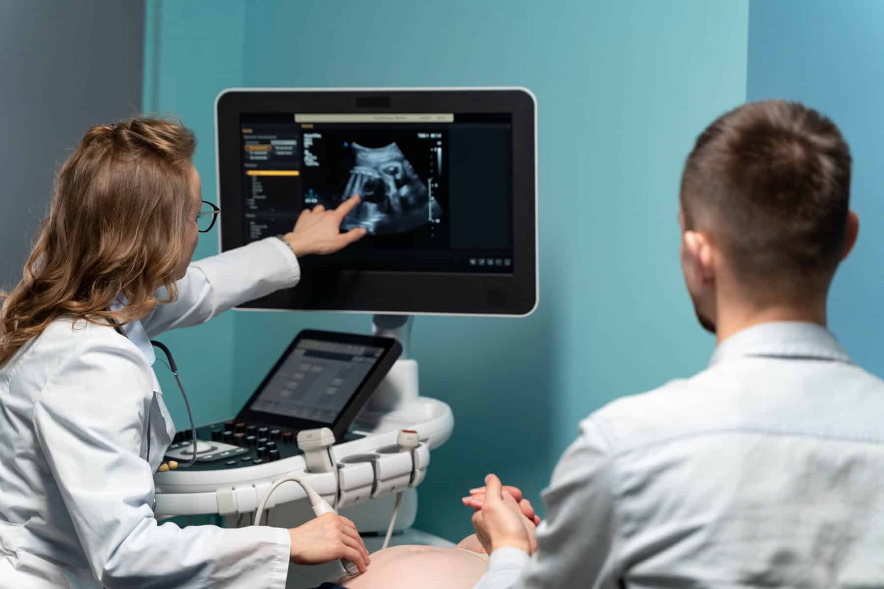
Ultrasound uses high-frequency sound waves to create images of organs and soft tissues inside the body. A handheld probe sends sound waves into the body. The echoes are reflected back to form real-time images on a screen. Unlike X-rays or CT scans, ultrasound does not use radiation. It is widely used for abdominal scans, pelvic ultrasound reports, thyroid imaging and many other applications because it is safe, non-invasive and effective.
For example:
- In an abdominal scan, the radiologist may assess the liver, gallbladder and kidneys for structural abnormalities.
- In a pelvic ultrasound, the uterus and ovaries are examined for cysts, fibroids or other changes.
- In a thyroid ultrasound, nodules are measured and assessed for their appearance and blood flow pattern.
Ultrasound reports can feel confusing because they use structured clinical language. Measurements and scan report terms are written for healthcare professionals. Without context, phrases such as “normal echotexture” or “mild enlargement” may seem unclear or concerning.
For instance: “Prostate measures 38 cc, mildly enlarged.”
This may sound alarming, but mild enlargement is extremely common in men over 50 and often relates to benign age-related change.
The Structure of an Ultrasound Report
A private ultrasound report UK clinics provide typically follows a standard format. Understanding the structure makes it easier to interpret.
Patient and Clinical Details
The report begins with your identifying details, the date of the scan and the reason it was requested. This section may mention symptoms such as abdominal pain or a palpable lump.
Example: “Clinical indication: Right upper quadrant pain. Query gallstones.” This simply explains why the scan was performed.
Findings Section
The findings section is the main body of the report. Each organ or area examined is listed separately. The sonographer or radiologist describes whether structures appear normal or whether any abnormalities are seen. For example, in abdominal ultrasound results, the liver, gallbladder, pancreas, kidneys and spleen are usually assessed individually.
Example of structured findings:
- Liver: Normal size. Homogeneous echotexture.
- Gallbladder: Contains a 7 mm echogenic focus with posterior acoustic shadowing.
- Common bile duct: 4 mm in diameter.
- Kidneys: Normal. No hydronephrosis.
Here, the “7 mm echogenic focus with acoustic shadowing” strongly suggests a gallstone. The 4 mm bile duct is within normal range, meaning there is no obvious blockage.
Impression or Conclusion
The impression or conclusion summarises the key findings. This is often the most important part for patients. It highlights whether the scan appears normal or identifies a specific issue. The clinician may suggest further imaging, blood tests or referral for specialist review.
Example: “Findings consistent with cholelithiasis. No evidence of biliary obstruction.” This means gallstones are present, but there is no blockage. Management depends on symptoms.

Common Terms You May See
Understanding common scan report terms can reduce anxiety and prevent misinterpretation.
“Unremarkable” does not mean the scan was unnecessary. It simply means the area examined appears within expected limits.
A cyst described as: “Anechoic, thin-walled, no internal septations” is typically simple and benign.
By contrast: “Complex cyst with internal echoes and septations” usually leads to further imaging.
Understanding Measurements
Ultrasound measurements explained in the report help determine whether structures fall within expected size ranges.
Size and Dimensions
Organs and abnormalities are measured in millimetres or centimetres.
Examples:
- Gallbladder wall thickness: Normal under 3 mm.
- Abdominal aorta: Normal under 3 cm.
- Ovarian cyst under 5 cm (premenopausal woman): Often monitored rather than treated.
A 2 mm kidney stone may pass naturally. A 12 mm stone may require intervention. Size changes management.
Volume Calculations
For certain organs, such as the prostate or thyroid gland, volume calculations are included.
Example: “Prostate volume 52 cc.” Normal adult prostate volume is typically under 30 cc.
52 cc indicates moderate enlargement, often linked to benign prostatic hyperplasia rather than cancer.
Blood Flow Assessment
Doppler ultrasound evaluates blood flow within vessels and organs.
Examples:
- Increased blood flow in the thyroid may suggest thyroiditis.
- Absent blood flow in a testicle can indicate torsion, which is an emergency.
- Increased vascularity within a mass may prompt further investigation.
Blood flow patterns often determine urgency.
When the Report Mentions Specific Conditions
Sometimes your private ultrasound report may reference specific conditions. Understanding how these appear on imaging can help clarify findings.
Gallstones
Gallstones typically appear as bright echoes within the gallbladder and cast a dark acoustic shadow behind them.
Example report wording: “Multiple mobile calculi seen within the gallbladder.”
Mobile means they move with position change, a typical benign gallstone feature.
Fibroids
Uterine fibroids are common non-cancerous growths in the womb.
Example: “Intramural fibroid measuring 2.8 cm within anterior uterine wall.”
Small fibroids like this are often incidental and monitored unless symptomatic.
Fatty Liver
When the report mentions increased echogenicity in the liver, it may suggest fatty infiltration.
Example: “Diffuse increased hepatic echogenicity in keeping with fatty infiltration.”
This often correlates with metabolic factors and may prompt blood tests and lifestyle advice.
What an Ultrasound Cannot Always Show
Ultrasound shows structure rather than function.
For example:
- It can confirm gallstones but cannot measure how effectively the gallbladder contracts.
- It can show a thyroid nodule but cannot confirm hormonal activity without blood tests.
- It can detect a cyst but cannot determine biochemical behaviour.
Very small abnormalities under 3–4 mm may not be clearly visualised. Early inflammatory changes can also appear subtle.
When to Seek Medical Advice
If your report recommends follow-up, referral or further imaging, it is important to act on this advice.
Example: “Recommend repeat scan in 6 months.”
This often applies to small thyroid nodules or ovarian cysts that require monitoring.
If your symptoms continue despite normal findings, further review is also important. For example, persistent abdominal pain with a normal ultrasound may require blood tests, endoscopy or CT imaging.
A normal private ultrasound does not always explain persistent pain or discomfort.
Use the Report as a Guide, Not a Diagnosis
A private ultrasound report UK patients receive provides valuable information, but it must be interpreted alongside your symptoms, medical history and clinical examination. The report guides next steps rather than delivering a complete diagnosis on its own.
For example, a “normal” scan in someone with ongoing symptoms does not invalidate those symptoms. It simply narrows the possibilities.
If you would like help understanding your private ultrasound results or arranging further assessment, contact The Forbury Clinic for expert review and clear, patient-focused advice.