Ultrasound is a safe, accessible and highly useful imaging method used across many areas of medicine. It is particularly useful in diagnosing conditions, guiding treatment and monitoring patient progress; all without the risks associated with radiation. Whether you’ve been referred for a scan or are exploring options for private ultrasound, you can feel reassured about its safety and effectiveness.
What Is an Ultrasound?
Ultrasound is a medical imaging technique that uses high-frequency sound waves to produce live images of internal body structures. These images help healthcare professionals examine soft tissues, organs and blood flow in real time.
Unlike X-rays, which use ionising radiation, or MRI scans, which use strong magnetic fields, ultrasound uses sound waves that bounce off internal structures. This makes it a much safer option, especially for pregnant women and patients who require repeat imaging. While X-rays are better suited for bones and MRIs for highly detailed soft tissue imaging, ultrasound stands out for being fast, accessible and radiation-free.
How Does an Ultrasound Work?
Ultrasound works by transmitting sound waves into the body and then collecting the echoes as they bounce back from tissues and organs. These echoes are used to form an image on a screen in real time.
Here’s how the process works:
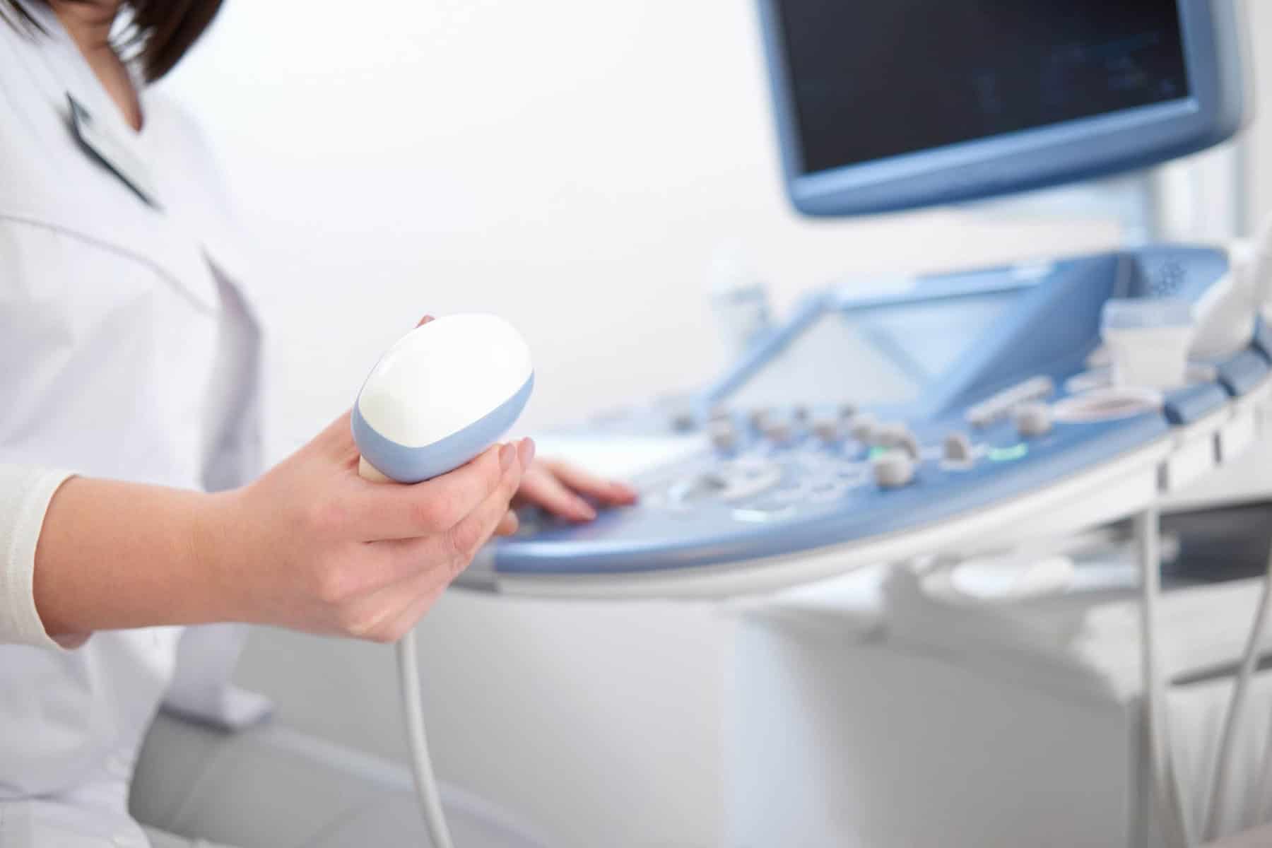
Sound wave emission: A small handheld device called a transducer is placed on the skin. It emits high-frequency sound waves that travel through the body.
Echo reception: As these sound waves hit tissues, fluids or organs, they bounce back to the transducer. Each type of tissue reflects sound differently, which is how different structures are displayed on the screen.
Image formation: The returning echoes are processed by a computer, which creates a real-time image displayed on a monitor. This allows the healthcare provider to see what’s happening inside the body as it happens.
As ultrasound uses real-time imaging, it’s particularly useful for observing movement — such as a beating heart or a developing baby in the womb.
Types of Ultrasound Scans
The Forbury Clinic provide several different types of ultrasound scans, each suited to specific parts of the body or types of investigation:
Abdominal ultrasound: Used to examine organs such as the liver, gallbladder, pancreas and kidneys. Often used to investigate abdominal pain, bloating or liver function concerns.
Bladder ultrasound: Examines the bladder and surrounding structures. It’s commonly used to assess bladder function, urinary retention or symptoms such as frequent urination.
Carotid ultrasound: Used to assess the carotid arteries in the neck, which supply blood to the brain and face. It helps detect narrowing or blockages that may increase the risk of stroke.
DVT ultrasound: This scan evaluates venous flow in the veins, typically in the legs, to detect deep vein thrombosis (DVT). Often used when symptoms like swelling or pain suggest a possible clot.
Kidney ultrasound: Captures detailed images of the kidneys and surrounding areas. Used to diagnose conditions such as kidney stones, cysts or suspected kidney disease.
Liver ultrasound: Assesses the liver’s size, texture and structure. Often used to detect liver disease, fatty liver, cysts or abnormal growths.
Lumps and Bumps ultrasound: Assesses soft tissue swellings or unexplained lumps under the skin. It helps determine whether a lump is solid, fluid-filled or potentially abnormal.
Musculoskeletal ultrasound: Used to examine joints, muscles, tendons and ligaments. Helpful in diagnosing injuries such as tendonitis, tears or inflammation.
Pelvic ultrasound: Used to examine organs within the pelvic area, such as the uterus, ovaries, bladder and prostate. Commonly used to investigate pelvic pain, abnormal bleeding or fertility concerns.
Testicle ultrasound: Also known as a scrotal ultrasound, this scan evaluates the testicles and surrounding tissues. It’s used to investigate pain, swelling, lumps or suspected torsion.
Thyroid ultrasound: Examines the thyroid gland in the neck to check for nodules, cysts or enlargement. It’s a key test in evaluating thyroid-related conditions.
Urinary Tract ultrasound: Designed to evaluate the kidneys, bladder and ureters, this ultrasound is often used to investigate symptoms such as urinary pain, recurrent infections or suspected kidney stones.
Uterine/ovary ultrasound: Provides detailed imaging of the reproductive organs, including the uterus and ovaries. Often used to investigate pelvic pain, bleeding or fertility issues.
What to Expect During an Ultrasound
If you’ve never had an ultrasound before, it’s natural to wonder what the experience will be like. Most scans are quick, painless and straightforward.
During the procedure:
- You’ll lie down in a comfortable position.
- A water-based gel will be applied to the area being examined. This gel helps the transducer move smoothly and improves sound wave transmission.
- The clinician will gently move the transducer across your skin to capture the necessary images.
- You may be asked to change position slightly or hold your breath briefly to improve image clarity.
Most ultrasound scans take between 15 and 45 minutes, depending on the area being examined. After the scan, you can return to your normal activities immediately.

Benefits of Ultrasound Imaging
Ultrasound offers several distinct advantages as a diagnostic tool:
No radiation: Safe for pregnant women and suitable for repeat use.
Real-time imaging: Allows clinicians to instantly see moving structures like heartbeat or blood flow.
Safe and non-invasive: No needles, incisions or recovery time needed.
Cost-effective: Generally more affordable than MRI or CT scans.
Limitations of Ultrasound
While ultrasound is a highly effective imaging method, it does have some limitations:
Less effective in areas with gas or dense bone: Structures like the lungs or bones can obstruct sound waves, making images less clear and difficult to assess.
May need follow-up imaging: In some cases, ultrasound provides enough information to make a diagnosis, but in others, a CT or MRI may be required for further clarity.
Image quality can vary: Results depend on the operator’s skill, the equipment quality and patient factors such as body type or movement. At Forbury clinic all the ultrasounds are performed by experience Consultant Radiologists.
If you have concerns about an upcoming ultrasound or would like to book a private ultrasound appointment at The Forbury Clinic, contact our team for help.
Ultrasound FAQs
How long does an ultrasound take?
Most ultrasound scans last between 15 and 45 minutes, depending on the complexity of the area being examined.
Is an ultrasound painful?
No, ultrasounds are generally painless. You may feel slight pressure from the transducer or the gel may feel cool on your skin, but the procedure is not uncomfortable.
Can I eat before an ultrasound?
It depends on the type of scan. For abdominal scans, you may be asked to fast beforehand. For other types, such as pelvic or musculoskeletal scans, you can usually eat as normal. Your healthcare provider will give you specific instructions.
What is the difference between 2D, 3D and Doppler ultrasound?
2D ultrasound provides flat, two-dimensional images and is the most common type.
3D ultrasound captures multiple 2D images to create a three-dimensional picture, often used in pregnancy scans.
Doppler ultrasound measures the direction and speed of blood flow in vessels, helping assess circulation and detect blockages.